LIMSwiki
目录
| 滑液關節 | |
|---|---|
 滑液關節的構造 | |
 | |
| 标识字符 | |
| 拉丁文 | junctura synovialis |
| TA98 | A03.0.00.020 |
| TA2 | 1533 |
| FMA | FMA:7501 |
| 《解剖學術語》 [在维基数据上编辑] | |
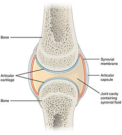
滑液關節(英語:Synovial joint)又稱滑膜关节,另稱動關節(英語:Diarthrosis),內部空間充有液體的關節,以纖維構成的關節囊連結相鄰的骨骼。關節囊延續接連骨骼的骨膜,構成滑液腔的外邊界並圍繞骨骼的關節表面。滑液腔充填有滑液。關節囊由外層(纖維膜)和內層(滑液膜)構成,外層維持骨與骨之間的連結結構,內層可製造與密封滑液[1][2]。
滑液關節是哺乳動物最常見且活動性最佳的關節類型[3]。與多數其他類型的關節一樣,滑液關節的活動位置在骨骼間的接觸點。
結構
滑液關節包含以下構造:
- 滑液腔:所有滑液關節在骨骼之間都有一個充填滑液的特有空間。
- 關節囊:延續自骨膜的纖維囊,包圍關節並連結組合該關節的骨骼。關節囊由兩層組成:(1)外層纖維膜,可包含韌帶;(2)內層滑液膜,分泌能潤滑、減震、滋養關節的滑液。關節囊富含神經,但沒有血管和淋巴管,是通過擴散(緩慢的過程)或對流(較高效率的過程,運動時產生)從周圍的血液供應中獲取營養。
- 關節軟骨:滑液關節內的骨骼被透明軟骨覆蓋,該層軟骨在骨骼末端的骨骺上襯有光滑的表面,使骨頭不會黏合在一起;關節軟骨的功能是吸收震動並減少動作過程中的摩擦。
許多(但不是全部)滑液關節也有以下額外的構造[4]:
- 關節盤或半月板:相對的關節面之間的纖維軟骨墊。
- 關節脂肪墊:保護關節軟骨的脂肪墊,比如膝關節的髕骨下脂肪墊。
- 肌腱:索狀的規則緻密結締組織,由平行的膠原蛋白束組成。
- 副韌帶(囊外和囊內):某些纖維膜的纖維排成規則緻密結締組織的平行束,適合抵抗拉力,防止極端動作造成關節損壞。
- 滑液囊:囊狀結構,充填與滑液類似的液體,可緩和某些關節(肩和膝)的摩擦。
血液供應
關節種類
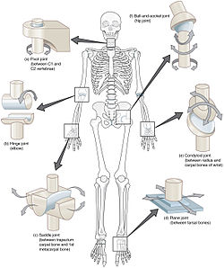
滑液關節有七種類型[5]。有些類型相對不易活動,但比較穩定。其他類型有多方向的活動自由度,但必須以受傷風險為代價[5]。按照活動性由低至高,依序是:
| 名稱 | 舉例 | 說明 |
|---|---|---|
| 滑動關節 | 腕骨、肩鎖關節 | 這些關節僅允許滑動,多軸運動如椎骨之間的關節[6]。 |
| 樞紐關節 | 肱尺關節 | 這些關節就像門的鉸鏈一樣,只允許在一個平面內屈曲和伸展。 |
| 樞軸關節 | 寰樞關節、近端橈尺關節、遠端橈尺關節 | 一個骨頭繞著另一骨頭旋轉,屬於單軸自由度[7]。 |
| 髁狀關節 橢球關節 |
橈腕關節 | 髁狀關節像是杵臼關節的改版,允許在兩個垂直軸內進行主要運動,第三軸向則是被動或輔助運動。某些分類法會將髁狀關節和橢球關節區分開來[8][9]。這些關節允許屈曲、伸展、外展、內收等動作(環行運動)。 |
| 鞍狀關節 | 腕掌關節[10][11]、胸鎖關節[12] | 關節相對的兩面呈凹形與凸形,如同鞍形,允許有與橢球關節類似但更大的活動度。 |
| 杵臼關節 | 肩關節[13]、髖關節 | 允許滑行以外所有方向的動作。 |
| 合成關節[14][15] 雙髁關節[16] |
膝關節 | 髁狀關節(股骨髁與脛骨髁相連)和鞍狀關節(股骨下端與髕骨相連)。 |
功能
滑液關節的動作可以有:
- 外展:遠離身體中線的動作。
- 內收:移近身體中線的動作。
- 伸展:在關節處伸直肢體。
- 屈曲:在關節處彎曲肢體。
- 旋轉:圍繞定點的圓周運動。
臨床意義
關節間隙等於關節內骨骼之間的距離。關節間隙變窄是骨關節炎和發炎性退化的徵兆[17]。正常的關節間隙在髖關節(上髖臼處)至少2mm[18],在膝關節至少3mm[19],在肩關節至少4到5mm[20],顳頜關節在1.5到4mm之間[21]。因此,關節間隙變窄的程度是骨關節炎的放射學分類要素之一。
參考文獻
- ^ Module - Introduction to Joints互联网档案馆的存檔,存档日期2008-05-26.
- ^ 存档副本. [2022-10-10]. (原始内容存档于2022-10-10).
- ^ Synovial Joints. Boundless Anatomy and Physiology. Lumen Learning. [2019-11-30]. (原始内容存档于2020-12-07).
- ^ Drake et al. (2009) Gray's Anatomy for Students, 2nd Edition, Skeletal system, p.21
- ^ 5.0 5.1 Umich 2010 couse, Module - Introduction to Joints (页面存档备份,存于互联网档案馆)
- ^ Moore, et al. Introduction to Clinically Oriented Anatomy. Baltimore: Lippincott Williams & Wilkins, 2006.
- ^ Platzer, Werner (2008) Color Atlas of Human Anatomy, Volume 1, p.28 (页面存档备份,存于互联网档案馆)
- ^ Rogers, Kara (2010) Bone and Muscle: Structure, Force, and Motion p.157 (页面存档备份,存于互联网档案馆)
- ^ Sharkey, John (2008) The Concise Book of Neuromuscular Therapy p.33 (页面存档备份,存于互联网档案馆)
- ^ Saddle joint - Definition, Movements, Examples and Diagrams. anatomy.co.uk. [2019-11-30]. (原始内容存档于2020-12-07).
- ^ Moore, KL. Clinically Oriented Anatomy 8. Philadelphia: Wolters Kluwer. 2018: 26. ISBN 9781496347213.
- ^ Moore, KL. Clinically Oriented Anatomy 8. Philadelphia: Wolters Kluwer. 2018: 264. ISBN 9781496347213.
- ^ And the phalanges (toes, fingers). Introduction to Joints: Synovial Joints - Ball and Socket Joints 互联网档案馆的存檔,存档日期2011-07-20.
- ^ Moini (2011) Introduction to Pathology for the Physical Therapist Assistant pp.231-2 (页面存档备份,存于互联网档案馆)
- ^ The Biophysical Foundations Of Human Movement (2005) By Bruce Abernethy pp.23, 331 (页面存档备份,存于互联网档案馆)
- ^ Drake, Richard L. (Richard Lee), 1950-. Gray's anatomy for students. Vogl, Wayne,, Mitchell, Adam W. M.,, Gray, Henry, 1825-1861. Third. Philadelphia, PA. : 20 [2019-11-29]. ISBN 9780702051319. OCLC 881508489. (原始内容存档于2021-03-02).
- ^ Jacobson, Jon A.; Girish, Gandikota; Jiang, Yebin; Sabb, Brian J. Radiographic Evaluation of Arthritis: Degenerative Joint Disease and Variations. Radiology. 2008, 248 (3): 737–747. ISSN 0033-8419. doi:10.1148/radiol.2483062112.
- ^ Lequesne, M. The normal hip joint space: variations in width, shape, and architecture on 223 pelvic radiographs. Annals of the Rheumatic Diseases. 2004, 63 (9): 1145–1151. ISSN 0003-4967. PMC 1755132
 . doi:10.1136/ard.2003.018424.
- ^ Page 6 in: Roland W. Moskowitz. Osteoarthritis: Diagnosis and Medical/surgical Management, LWW Doody's all reviewed collection. Lippincott Williams & Wilkins. 2007. ISBN 9780781767071.
- ^ Glenohumeral joint space. radref.org. [2019-11-29]. (原始内容存档于2020-12-07)., in turn citing: Petersson, Claes J.; Redlund-Johnell, Inga. Joint Space in Normal Gleno-Humeral Radiographs. Acta Orthopaedica Scandinavica. 2009, 54 (2): 274–276. ISSN 0001-6470. doi:10.3109/17453678308996569.
- ^ Massilla Mani, F.; Sivasubramanian, S. Satha. A study of temporomandibular joint osteoarthritis using computed tomographic imaging. Biomedical Journal. 2016, 39 (3): 201–206. ISSN 2319-4170. PMC 6138784
 . doi:10.1016/j.bj.2016.06.003.
參考書目
- 《人體學習百科》,貓頭鷹出版社,ISBN 957-9684-11-1


