Type a search term to find related articles by LIMS subject matter experts gathered from the most trusted and dynamic collaboration tools in the laboratory informatics industry.
| Torticollis | |
|---|---|
| Other names |
|
 | |
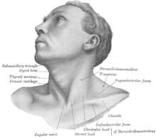
| The muscles involved with torticollis | |
| Specialty | Orthopedics |
| Diagnostic method | Ultrasonography |
Torticollis, also known as wry neck, is a painful, dystonic condition defined by an abnormal, asymmetrical head or neck position, which may be due to a variety of causes. The term torticollis is derived from Latin tortus 'twisted' and collum 'neck'.[1][2]
The most common case has no obvious cause, and the pain and difficulty in turning the head usually goes away after a few days, even without treatment in adults.
Torticollis is a fixed or dynamic tilt, rotation, with flexion or extension of the head and/or neck.
The type of torticollis can be described depending on the positions of the head and neck.[1][3][4]
A combination of these movements may often be observed. Torticollis can be a disorder in itself as well as a symptom in other conditions.
Other signs and symptoms include:[8][9]
A multitude of conditions may lead to the development of torticollis including: muscular fibrosis, congenital spine abnormalities, or toxic or traumatic brain injury.[2] A rough categorization discerns between congenital torticollis and acquired torticollis.[10]
Other categories include:[11]
Congenital muscular torticollis is the most common torticollis that is present at birth.[12] Congenital muscular torticollis is the third most common congenital musculoskeletal deformity in children.[13] The cause of congenital muscular torticollis is unclear. Birth trauma or intrauterine malposition is considered to be the cause of damage to the sternocleidomastoid muscle in the neck.[2] Other alterations to the muscle tissue arise from repetitive microtrauma within the womb or a sudden change in the calcium concentration in the body that causes a prolonged period of muscle contraction.[14]
Any of these mechanisms can result in a shortening or excessive contraction of the sternocleidomastoid muscle, which curtails its range of motion in both rotation and lateral bending. The head is typically tilted in lateral bending toward the affected muscle and rotated toward the opposite side. In other words, the head itself is tilted in the direction of the shortened muscle, with the chin tilted in the opposite direction.[11]
Congenital torticollis is presented at 1–4 weeks of age, and a hard mass usually develops. It is normally diagnosed using ultrasonography and a color histogram or clinically by evaluating the infant's passive cervical range of motion.[15]
Congenital torticollis constitutes the majority of cases seen in paediatric clinical practice.[11] The reported incidence of congenital torticollis is 0.3-2.0%.[16] Sometimes a mass, such as a sternocleidomastoid tumor, is noted in the affected muscle. Congenital Muscular Torticollis is also defined by a fibrosis contracture of the sternocleidomastoid muscle on one side of the neck.[13] Congenital torticollis may not resolve on its own, and can result in rare complications including plagiocephaly.[17] Secondary complications associated with Congenital Muscular Torticollis include visual dysfunctions, facial asymmetry, delayed development, cervical scoliosis, and vertebral wedge degeneration which will have a serious impact on the child's appearance and even mental health.[13]
Benign paroxysmal torticollis is a rare disorder affecting infants. Recurrent attacks may last up to a week. The condition improves by age 2. The cause is thought to be genetic.[18]
Noncongenital muscular torticollis may result from muscle spasm, trauma, scarring or disease of cervical vertebrae, adenitis, tonsillitis, rheumatism, enlarged cervical glands, retropharyngeal abscess, or cerebellar tumors.[19] It may be spasmodic (clonic) or permanent (tonic). The latter type may be due to Pott's Disease (tuberculosis of the spine).[20]
Most commonly this self-limiting form relates to an untreated dental occlusal dysfunction, which is brought on by clenching and grinding the teeth during sleep. Once the occlusion is treated it will completely resolve. Treatment is accomplished with an occlusal appliance, and equilibration of the dentition.
Torticollis with recurrent, but transient contraction of the muscles of the neck and especially of the sternocleidomastoid, is called spasmodic torticollis. Synonyms are "intermittent torticollis", "cervical dystonia" or "idiopathic cervical dystonia", depending on cause.[23]
Torticollis can be caused by damage to the trochlear nerve (fourth cranial nerve), which supplies the superior oblique muscle of the eye. The superior oblique muscle is involved in depression, abduction, and intorsion of the eye. When the trochlear nerve is damaged, the eye is extorted because the superior oblique is not functioning. The affected person will have vision problems unless they turn their head away from the side that is affected, causing intorsion of the eye and balancing out the extorsion of the eye. This can be diagnosed by the Bielschowsky test, also called the head-tilt test, where the head is turned to the affected side. A positive test occurs when the affected eye elevates, seeming to float up.[24]
The main job of the sternocleidomastoid muscle is to help move the head and neck by turning the head to one side and bending the neck forward.[25] The sternocleidomastoid muscle gets its blood from different arteries in the neck, which bring oxygen and nutrients to keep the muscle healthy. Torticollis can happen when there are issues with the sternocleidomastoid muscle, like if it's too short, causing the head and neck to be in an odd position.[25] Torticollis can also be caused by problems with bones, muscles, or the spine in the neck, leading to difficulty moving the head and neck normally.[25] Knowing about the sternocleidomastoid muscle and how it works is crucial for doctors to diagnose and treat torticollis correctly, so they can find and fix the problem causing it. Differences in how the sternocleidomastoid muscle is supplied with blood or nerves can affect how torticollis develops or how well treatments work, so it's important for doctors to consider these variations when planning treatment.[26] Having a good understanding of the neck's anatomy helps doctors accurately diagnose torticollis and choose the best treatments to help patients feel better.
The sternocleidomastoid muscle gets signals from nerves in the neck and head to contract and move properly. The underlying anatomical distortion causing torticollis is a shortened sternocleidomastoid muscle. This is the muscle of the neck that originates at the sternum and clavicle and inserts on the mastoid process of the temporal bone on the same side.[11] There are two sternocleidomastoid muscles in the human body and when they both contract, the neck is flexed. The main blood supply for these muscles come from the occipital artery, superior thyroid artery, transverse scapular artery and transverse cervical artery.[11] The main innervation to these muscles is from cranial nerve XI (the accessory nerve) but the second, third and fourth cervical nerves are also involved.[11] Pathologies in these blood and nerve supplies can lead to torticollis.[citation needed]
Evaluation of a child with torticollis begins with history taking to determine circumstances surrounding birth and any possibility of trauma or associated symptoms. Physical examination reveals decreased rotation and bending to the side opposite from the affected muscle. Some[who?] say that congenital cases more often involve the right side, but there is not complete agreement about this in published studies. Evaluation should include a thorough neurologic examination, and the possibility of associated conditions such as developmental dysplasia of the hip and clubfoot should be examined. Radiographs of the cervical spine should be obtained to rule out obvious bony abnormality, and MRI should be considered if there is concern about structural problems or other conditions.
Ultrasonography can be used to visualize muscle tissue, with a colour histogram generated to determine cross-sectional area and thickness of the muscle.[27]
Evaluation by an optometrist or an ophthalmologist should be considered in children to ensure that the torticollis is not caused by vision problems (IV cranial nerve palsy, nystagmus-associated "null position", etc.).
Differential diagnosis for torticollis includes[11][28]
Cervical dystonia appearing in adulthood has been believed to be idiopathic in nature, as specific imaging techniques most often find no specific cause.[30]
Teaching people how to sit and stand properly can help reduce strain on the neck muscles and improve posture. Changing habits like bad posture or repetitive movements can help ease symptoms of torticollis.[26] Wearing a special collar can also support the neck and keep it in the right position during daily activities. Using electrical devices have also been shown to reduce pain, make muscles work better, and relax tight muscles.[31] Injecting a substance like Botox into overactive muscles can weaken them temporarily, allowing for better movement.[32] If other treatments don't work, surgery might be needed to fix the muscles or bones causing torticollis.
Physical therapy is an option for treating torticollis in a non-invasive and cost-effective manner.[33] In the children above 1 year of age, surgical release of the tight sternocleidomastoid muscle is indicated along with aggressive therapy and appropriate splinting. Occupational therapy rehabilitation in congenital muscular torticollis concentrates on observation, orthosis, gentle stretching, myofascial release techniques, parents’ counseling-training, and home exercise program. While outpatient infant physiotherapy is effective, home therapy performed by a parent or guardian is just as effective in reversing the effects of congenital torticollis.[14] It is important for physical therapists to educate parents on the importance of their role in the treatment and to create a home treatment plan together with them for the best results for their child.
Five components have been recognized as the "first choice intervention" in PT for treatment of torticollis and include
In therapy, parents or guardians should expect their child to be provided with these important components, explained in detail below.[34] Lateral neck flexion and overall range of motion can be regained quicker in newborns when parents conduct physical therapy exercises several times a day.[14]
Physical therapists should teach parents and guardians to perform the following exercises:[14]
Physical therapists often encourage parents and caregivers of children with torticollis to modify the environment to improve neck movements and position. Modifications may include:
Environmental Modifications for Torticollis Management:
A meta-analysis shows physical therapists specializing in manual therapy have developed effective interventions for the management of Congenital Muscular Torticollis (CMT), primarily centered around massage and passive stretching techniques. These interventions are tailored to address the specific needs of pediatric patients, with a focus on stretching the sternocleidomastoid muscle.[36] Various protocols have been proposed, including stretching exercises held for specific durations and repetitions, aimed at increasing blood flow, and promoting muscle relaxation.
Additionally, massage maneuvers such as rhythmic muscle mobilization techniques are employed to mobilize cervical structures and induce relaxation.[36] The systematic review highlights the efficacy of manual therapy and passive stretching in improving cervical range of motion (ROM) in children with CMT. Furthermore, the involvement of caregivers in home exercise programs is emphasized as crucial for optimizing treatment outcomes and promoting motor development while preventing secondary complications.
A systematic review, looked into the possible benefits of using manipulation techniques to counteract infant torticollis. The study considered the impact of manipulation on an infant's sleep, crying, and restlessness as well.[37] This review did not report any adverse effects of using manipulation techniques. It was shown that using manipulation techniques on their own had little to no statistical differences from a placebo group, immediately. When manipulation techniques were combined with physical therapy, there was a change in symptoms compared to the use of physical therapy alone. When targeting the cervical spine, manipulation techniques were shown to shorten treatment duration in infants with head asymmetries.[37]
A Korean study has recently[when?] introduced an additional treatment called microcurrent therapy that may be effective in treating congenital torticollis. For this therapy to be effective the children should be under three months of age and have torticollis involving the entire sternocleidomastoid muscle with a palpable mass and a muscle thickness over 10 mm. Microcurrent therapy sends minute electrical signals into tissue to restore the normal frequencies in cells.[27] Microcurrent therapy is completely painless and children can only feel the probe from the machine on their skin.[27]
Microcurrent therapy is thought to increase ATP and protein synthesis as well as enhance blood flow, reduce muscle spasms and decrease pain along with inflammation.[27] It should be used in addition to regular stretching exercises and ultrasound diathermy. Ultrasound diathermy generates heat deep within body tissues to help with contractures, pain and muscle spasms as well as decrease inflammation. This combination of treatments shows remarkable outcomes in the duration of time children are kept in rehabilitation programs: Micocurrent therapy can cut the length of a rehabilitation program almost in half with a full recovery seen after 2.6 months.[27]
About 5–10% of cases fail to respond to stretching and require surgical release of the muscle.[38][39]
Surgical release involves the two heads of the sternocleidomastoid muscle being dissected free. This surgery can be minimally invasive and done laparoscopically. Usually surgery is performed on those who are over 12 months old. The surgery is for those who do not respond to physical therapy or botulinum toxin injection or have a very fibrotic sternocleidomastoid muscle.[8] After surgery the child will be required to wear a soft neck collar (also called a Callot's cast). There will be an intense physiotherapy program for 3–4 months as well as strengthening exercises for the neck muscles.[40]
Other treatments include:[14]
CMT is a neck problem that babies are born with or develop soon after birth, causing their neck to be stiff and bent in an awkward position.[36] Besides the sternocleidomastoid muscle, other muscles in the neck can also be affected by CMT, leading to problems moving the head and neck normally.[36] The main goal of treating CMT is to make the sternocleidomastoid muscle stronger and more flexible, so the neck can move better and symptoms can improve.
Studies and evidence from clinical practice show that 85–90% of cases of congenital torticollis are resolved with conservative treatment such as physical therapy.[34] Earlier intervention is shown to be more effective and faster than later treatments. More than 98% of infants with torticollis treated before 1 month of age recover by 2.5 months of age.[34] Infants between 1 and 6 months usually require about 6 months of treatment.[34] After that point, therapy will take closer to 9 months, and it is less likely that the torticollis will be fully resolved.[34] It is possible that torticollis will resolve spontaneously, but chance of relapse is possible.[11] For this reason, infants should be reassessed by their physical therapist or other provider 3–12 months after their symptoms have resolved.[34]

In veterinary literature usually only the lateral bend of head and neck is termed torticollis, whereas the analogon to the rotatory torticollis in humans is called a head tilt. The most frequently encountered form of torticollis in domestic pets is the head tilt, but occasionally a lateral bend of the head and neck to one side is encountered.[43]
Causes for a head tilt in domestic animals are either diseases of the central or peripheral vestibular system or relieving posture due to neck pain. Known causes for head tilt in domestic animals include: